
The Day Medicine's Greatest Wall Finally Came Down
There has always been a limit that medicine could not cross. Not a limit of effort or intelligence, but a biological one, written into the very nature of living things. Once cells die, they do not come back. Once an organ is damaged beyond repair, it stays that way. This has been one of the most fundamental rules of biology for as long as medicine has existed as a discipline.
The consequences of that rule are familiar to anyone who has watched a loved one live with its effects. A stroke destroys neurons in the brain, and the movement those neurons once controlled does not return. A heart attack kills cardiac muscle cells, and the heart pumps less effectively for the rest of a patient’s life. A spinal cord injury severs the pathways that carry signals to the limbs, and what is severed stays severed. What medicine has been able to offer in these situations is real and often valuable: drugs that support remaining function, prosthetic limbs, artificial hearts, dialysis machines, rehabilitation programmes designed to help patients compensate for what has been lost. But the lost tissue itself has remained lost. The best medicine could do was manage the damage, not undo it.
On February 19, 2026, Japan’s Ministry of Health, Labour and Welfare approved the manufacture and sale of two regenerative medicine products derived from induced pluripotent stem cells, or iPS cells. The two diseases targeted are Parkinson’s disease and severe heart failure, both conditions that have long been considered beyond the reach of genuinely curative treatment. These approvals are expected to represent the world’s first regulatory approval of iPS cell-based regenerative medicine products, and they mark the moment when a technology capable of actually replacing lost cells, rather than simply compensating for their absence, crossed the threshold from experimental science into clinical practice.
The scientific roots of this moment go back to 2006, when Professor Shinya Yamanaka of Kyoto University succeeded in generating iPS cells from mouse tissue. He received the Nobel Prize in Physiology or Medicine in 2012 for that work. Nearly twenty years of intensive research, regulatory development, and clinical testing have followed. What emerged from a research laboratory two decades ago is now something a physician can prescribe.
Planting New Neurons in the Brain: Amscepri for Parkinson's Disease
The first of the two newly approved products is Amscepri, known by its generic name ragneprocell, developed by Sumitomo Pharma. It is designed for patients with advanced Parkinson’s disease who no longer respond adequately to existing drug treatments.
To understand why this matters, it helps to understand what Parkinson’s disease actually does to the brain. Deep within the brain lies a region called the substantia nigra, a cluster of neurons whose job is to produce dopamine, the chemical messenger that plays a central role in coordinating smooth, controlled movement. In Parkinson’s disease, these neurons gradually die. As dopamine levels fall, the signals that normally flow between the brain and the body become disrupted. The characteristic symptoms follow: tremor at rest, rigidity in the muscles, a slow and shuffling gait, difficulty initiating movement. In Japan alone, roughly 200,000 people are living with the disease, and the number is rising as the population ages.
The standard treatment for Parkinson’s disease has for decades been a drug called levodopa, commonly written as L-dopa. Levodopa is converted into dopamine in the brain, partially replacing what the dying neurons can no longer produce. For many patients, especially in the early stages of the disease, levodopa works reasonably well. But as the disease progresses and more neurons are lost, the drug becomes increasingly unreliable. Patients begin to experience what is called the wearing-off phenomenon, in which the therapeutic effect of each dose fades before the next one is due, leaving them in states of severely impaired movement for hours at a time. On the other side of the balance, when the drug is working too strongly, it can cause dyskinesia, which means uncontrolled and sometimes violent involuntary movements. Managing advanced Parkinson’s disease with levodopa often feels like trying to walk a tightrope, with debilitating symptoms waiting on both sides.
Amscepri takes a fundamentally different approach. Rather than supplementing a depleted chemical, it attempts to restore the cellular source of that chemical. The product consists of dopaminergic neural progenitor cells, which are immature nerve cells destined to develop into the same type of dopamine-producing neurons that Parkinson’s disease destroys. These cells are derived not from the patient’s own body but from iPS cells prepared in advance from donor material. During the procedure, the cells are injected directly into a region of the brain called the putamen, using a form of precision neurosurgery called stereotactic surgery. Once in place, the transplanted cells mature within the brain environment and begin producing dopamine on their own, continuously and without the peaks and valleys associated with drug dosing.
The clinical trial results, while based on a small number of patients, were striking. Six patients received the transplant, and four showed measurable improvement in motor function. The most compelling evidence came from PET imaging, a brain scanning technique that can visualise the presence and activity of dopamine in specific regions. Before transplantation, the affected areas of the brain appeared dark in the scans, indicating the near-total absence of dopamine activity. One year after the procedure, those same areas showed clear signals of dopamine presence and function. Living, working neurons had taken root in the brain and were doing the job that the patient’s own cells could no longer do.
A Sheet That Beats Like a Heart: ReHeart for Severe Heart Failure
The second approved product, ReHeart, was developed by Cuorips, a bioventure company that grew out of research at Osaka University led by Professor Emeritus Yoshiki Sawa. Its target is ischemic cardiomyopathy, a form of severe heart failure caused by damage to the heart muscle following events such as a heart attack.
Heart failure, at its most basic, is a failure of the heart to function as a pump. The heart’s job is to contract rhythmically and push blood out to the rest of the body, and it does this by means of specialised muscle cells called cardiomyocytes. When a heart attack occurs, part of the blood supply to the heart is cut off. The cardiomyocytes in the affected area are deprived of oxygen and begin to die. Unlike many other cell types in the body, heart muscle cells have a very limited ability to regenerate on their own, which means the dead tissue is replaced by scar tissue rather than functional muscle. The heart that remains is weaker and less able to maintain adequate circulation.
In the later stages of severe heart failure, the medical options narrow dramatically. Heart transplantation is the definitive treatment, but donor hearts are desperately scarce. In Japan, the gap between patients who need a transplant and the number of donated hearts available is so wide that many patients wait years and do not survive long enough to receive one. The alternative is a ventricular assist device, a mechanical pump that either supplements or takes over the heart’s pumping function. These devices extend life and can be effective, but they require either a partially implanted device connected to an external power source or a fully implanted system, both of which impose significant constraints on daily life. Patients cannot drive. Infection risk is elevated. The psychological burden of living with a machine performing the work of your heart is substantial, and it is a burden that can last for years while a patient waits for a transplant that may never come.
ReHeart works differently. iPS cells are guided through a specialised laboratory process to differentiate into cardiomyocytes, and those cells are then formed into thin, flexible sheets. These sheets are not inert. When maintained in culture, they beat spontaneously, contracting and relaxing just as heart tissue does inside the body. They are sheets of living, active heart muscle.
Three of these beating sheets are applied directly to the surface of the patient’s weakened heart during surgery. The mechanism of benefit is not primarily that the sheets take over the pumping function directly, but rather that they release a range of signalling proteins called cytokines that stimulate the formation of new blood vessels in the surrounding tissue and reactivate dormant cardiomyocytes that have been suppressed by the disease environment. The effect is a gradual improvement in the contractile strength of the heart as a whole. In the clinical trial, four of the eight patients who received the treatment showed meaningful improvement in measures of cardiac function, including peak oxygen uptake, which is a reliable indicator of how well the heart is supplying the body during physical activity.
What iPS Cells Actually Are: Winding the Biological Clock Back Inside a Cell
To fully appreciate what has been achieved, it is worth taking a moment to understand what iPS cells are and why their existence was once considered close to impossible.
The human body is built from roughly 37 trillion cells. Skin cells, liver cells, neurons, cardiomyocytes, immune cells: they are all radically different from one another in their structure and function, yet they all contain exactly the same DNA. The reason they look and behave so differently is not that their genetic material differs, but that different sets of genes are switched on or off in each cell type. A skin cell has silenced the genes relevant to heart function. A neuron has silenced the genes relevant to digestion. This process of a cell committing to a particular identity and suppressing all other possibilities is called differentiation.
For most of the history of biology, differentiation was understood to be a one-way journey. Cells move from an undifferentiated state toward a specialised one as an organism develops, and that direction of travel was thought to be irreversible. A skin cell could not become a neuron. A cardiomyocyte could not become a liver cell. The analogy often used is a stone rolling down a hillside: once it reaches the bottom, it cannot roll back up on its own.
What Yamanaka demonstrated in 2006 was that this hillside could, in fact, be climbed. By introducing just four specific genes into a mature skin cell, which are now known worldwide as the Yamanaka factors and technically named Oct3/4, Sox2, Klf4, and c-Myc, he found that the cell could be forced to abandon its specialised identity and revert to a state resembling that of a very early embryo. The cell, in effect, forgot what it had become. From that reverted state, it could be guided back through differentiation toward any cell type the researcher chose: neurons, cardiomyocytes, liver cells, pancreatic cells, bone cells, blood cells. The possibilities extend across virtually the full range of cells that make up the human body.
The resulting cells are called induced pluripotent stem cells because they are induced, meaning artificially created, and pluripotent, meaning capable of giving rise to many different cell types. Their two defining properties are what make them medically valuable. The first is pluripotency itself, the ability to become virtually any cell type in the body. The second is self-renewal: in appropriate culture conditions, iPS cells can be maintained and multiplied essentially indefinitely, providing an unlimited supply of starting material. Together, these two properties make it possible to do something that was previously impossible: to manufacture the specific cell type that a patient’s body has lost, in whatever quantity is needed, and to deliver it to the site of damage.
Two Walls That Once Seemed Immovable, and How They Fell
Before iPS cells existed, the leading candidate for regenerative medicine was a different type of pluripotent cell called an embryonic stem cell, or ES cell. ES cells share many of the same remarkable properties as iPS cells, but the method of obtaining them created two problems that proved extremely difficult to resolve.
The first was ethical. ES cells are derived from human embryos at a very early stage of development, a process that destroys the embryo. In many countries and across many religious and philosophical traditions, this was regarded as a serious ethical violation involving the destruction of a potential human life. The controversy was not merely theoretical. During the administration of President George W. Bush in the United States, federal funding for research involving new human ES cell lines was prohibited, effectively halting a significant portion of American research in the field. Laboratories around the world navigated a patchwork of national regulations, some permissive and some highly restrictive, that complicated international collaboration and slowed progress. In Europe, individual countries took sharply different positions, making coordinated research across borders difficult.
The second problem was immunological. ES cells derived from an embryo carry the genetic identity of whoever donated that embryo, not of the patient who will receive the treatment. The patient’s immune system, which is finely tuned to distinguish between the body’s own cells and foreign material, identifies ES cell-derived tissue as foreign and mounts an attack against it. To prevent rejection, patients would need to take powerful immunosuppressive drugs indefinitely, broadly suppressing immune function and leaving them more vulnerable to infections and certain types of cancer.
iPS cells resolved both problems simultaneously. Because they can be created from the patient’s own skin or blood cells, no embryo is involved, and the ethical objection disappears. Because the cells carry the patient’s own genetic identity, the immune system does not recognise them as foreign, and rejection does not occur. When Yamanaka’s results were published in 2006, researchers around the world grasped immediately that the fundamental barriers to pluripotent stem cell research had been lifted. The pace of work accelerated sharply, and within a few years iPS cell research had become one of the most intensively pursued fields in all of biomedical science.
The Cost Problem: How Japan Cracked the Hundred-Million-Yen Barrier
Even after the scientific and ethical hurdles had been cleared, a severe practical obstacle remained. Making iPS cells is not simple, and producing them for individual patients at clinical quality is extraordinarily expensive.
The original vision for iPS cell medicine was fully personalised treatment. A patient’s own cells would be taken, reprogrammed into iPS cells, differentiated into whatever cell type was needed, tested to confirm quality and safety, and then transplanted back. Because the cells came from the patient themselves, there would be no immune rejection at all. This approach, called autologous transplantation, was regarded in the early years of the technology as the ideal endpoint.
The problem is time and money. Creating a clinical-grade iPS cell line from scratch for a single patient takes several months from start to finish. Cost estimates for the full process have ranged from tens of millions to over 100 million yen per patient. For a person experiencing acute heart failure, a wait of several months is not clinically acceptable. For a healthcare system trying to serve large numbers of patients, treatment costs at that level cannot be sustained within any realistic public reimbursement framework.
Japan’s response to this challenge was a national programme called the iPS Cell Stock Project. Rather than making cells for each patient individually, the project creates a library of iPS cell lines in advance, derived from carefully selected donors. The cells are grown at scale, quality-tested, and then frozen and stored, ready to be dispatched to hospitals on demand, much like a pharmaceutical product already in stock.
The key to making this work is the immune system’s recognition mechanism. The immune system identifies foreign cells by examining proteins on their surface called HLA molecules, which function as a kind of biological identity card. When HLA molecules from a donor cell match those of the recipient closely enough, the immune system accepts the transplanted tissue rather than attacking it. The statistical challenge is that a close HLA match between two unrelated individuals occurs, on average, only once in many thousands of random pairings.
The iPS Cell Stock Project found an elegant solution. By selecting donors who carry HLA types that are particularly common in the Japanese population, and who are homozygous for those types, meaning they inherited the same HLA variant from both parents, it becomes possible to cover a large fraction of the population with a small number of donor cell lines. Research has shown that iPS cell lines from just four carefully chosen homozygous donors can provide a sufficiently close HLA match for approximately 40 percent of the Japanese population. The cells in the stock can be dispatched immediately when a patient needs them, the cost per treatment falls dramatically compared to the personalised approach, and the two newly approved products, Amscepri and ReHeart, are both built on this stockpile model.
Research into the next generation of the technology is already well under way. Scientists are using gene editing tools, including CRISPR-Cas9, to delete the HLA class I molecules from iPS cells entirely, making those cells essentially invisible to the immune system. A cell that the immune system cannot recognise cannot be rejected. If this approach reaches clinical maturity, the result would be what researchers are calling universal iPS cells: a single cell line that could be transplanted into any patient anywhere in the world without triggering rejection, regardless of their genetic background. The social implication of this is sometimes described as the democratisation of regenerative medicine, the possibility that this form of treatment could become accessible to any patient, in any country, without the constraints of immune matching or the cost of personalised manufacturing.
From Keeping People Alive to Keeping People Well: What This Means for Society by 2050
The significance of these approvals extends well beyond the immediate benefit to patients with Parkinson’s disease or heart failure. If regenerative medicine fulfils the potential that its scientific foundations suggest, it will alter some of the most basic assumptions on which modern societies are built.
The global market for regenerative and cell-gene therapies is projected to reach the equivalent of approximately 200 trillion yen by 2050. But the more profound shift is not economic. It is a change in what medicine is fundamentally designed to do.
For most of medical history, the central task of medicine has been to prevent death: to keep damaged bodies functioning long enough for patients to live out a natural lifespan. Regenerative medicine makes it possible to pursue a different goal, not merely keeping the body going despite its injuries, but restoring it to genuine functional health. Lost neurons replaced. Damaged heart muscle repaired. The body not sustained around its damage but healed through it.
If that vision is realised at scale, the social structures built around the assumption that ageing necessarily means functional decline will face significant pressure. The concept of a mandatory retirement age was shaped in part by the practical reality that physical and cognitive capacity tend to diminish with time. If neurodegenerative diseases can be treated by replacing lost neurons, and cardiovascular decline reversed by restoring heart muscle, the relationship between age and capability may look very different a generation from now. Social security systems that were designed around a relatively short period of dependency at the end of life may need to be fundamentally reconceived. The notion that people have one career, one productive phase, and one period of decline could give way to a more fluid model in which biological restoration makes repeated reinvention genuinely possible.
Japan’s strategic position in this field is worth noting. Kyoto University holds core patents on iPS cell technology, and Japan has invested heavily in building the regulatory and manufacturing infrastructure that clinical translation requires. Some foundational patents are expected to expire in December 2026, which is likely to accelerate both competition from other companies and cost reduction for patients. Whether Japan can maintain its leading position will depend in large part on its ability to accumulate further clinical evidence and to shape the global regulatory standards that will govern how iPS cell medicines are evaluated, approved, and reimbursed worldwide.
Not a Finish Line, but a Starting Point
Professor Yoshiki Sawa of Osaka University, one of the key figures behind ReHeart, has described today’s approval not as a goal but as a waypoint. That framing is scientifically accurate and worth keeping in mind.
Both products were approved under a Japanese regulatory mechanism called conditional and time-limited approval, which is designed specifically for advanced therapeutic products targeting serious conditions where conventional evidence generation would take too long given patients’ urgent needs. Under this framework, approval is granted on the basis of preliminary evidence that a treatment is reasonably likely to be effective and has an acceptable safety profile. The manufacturer is then required to conduct confirmatory studies within a defined time period, with continued authorisation dependent on the results. Amscepri must generate confirmatory evidence within seven years of approval. The trials that supported these applications involved six patients for Amscepri and eight for ReHeart. These are not large numbers. Questions about the long-term durability of transplanted cells, the stability of the therapeutic effect over many years, the potential for the transplanted cells to behave in unexpected ways over time, and the full range of possible side effects will only be answered as more patients are treated and followed over longer periods.
None of this diminishes the significance of what has been achieved. The journey that led here began with Yamanaka running marathons to raise public donations for his research at a time when iPS cell science was still struggling to attract the funding it needed. It continued through years of painstaking progress in understanding how to guide pluripotent cells reliably toward specific fates, and in developing the manufacturing and quality-control systems that clinical-grade production demands. It required the creation of an entirely new regulatory framework capable of handling biological products that had no precedent in the history of drug approval.
The assumption that once-lost tissue is permanently gone has shaped clinical medicine throughout its entire history. Every treatment protocol, every rehabilitation approach, every decision about how to care for patients with neurological or cardiac disease has been built on top of that assumption. What these approvals announce, carefully and with appropriate scientific caution, is that the assumption may no longer hold in the way it once did.
The cells placed in patients’ brains and hearts during the trials that led to these approvals were living. They took root. They produced dopamine. They released signals that helped failing hearts begin to recover strength. Twenty years after a researcher in Kyoto demonstrated that the biological clock inside a cell could be wound back, that discovery is functioning in human bodies as approved medicine.
If damaged and lost parts of the body can gradually be restored, the question that follows is not only a medical one. It is a question about what kind of lives people will lead with the longer and more functional time they are given, and what kind of societies we will need to build to support them. Regenerative medicine has placed that question on the table. Working out the answers will take considerably longer than the science did.
Reference Links:
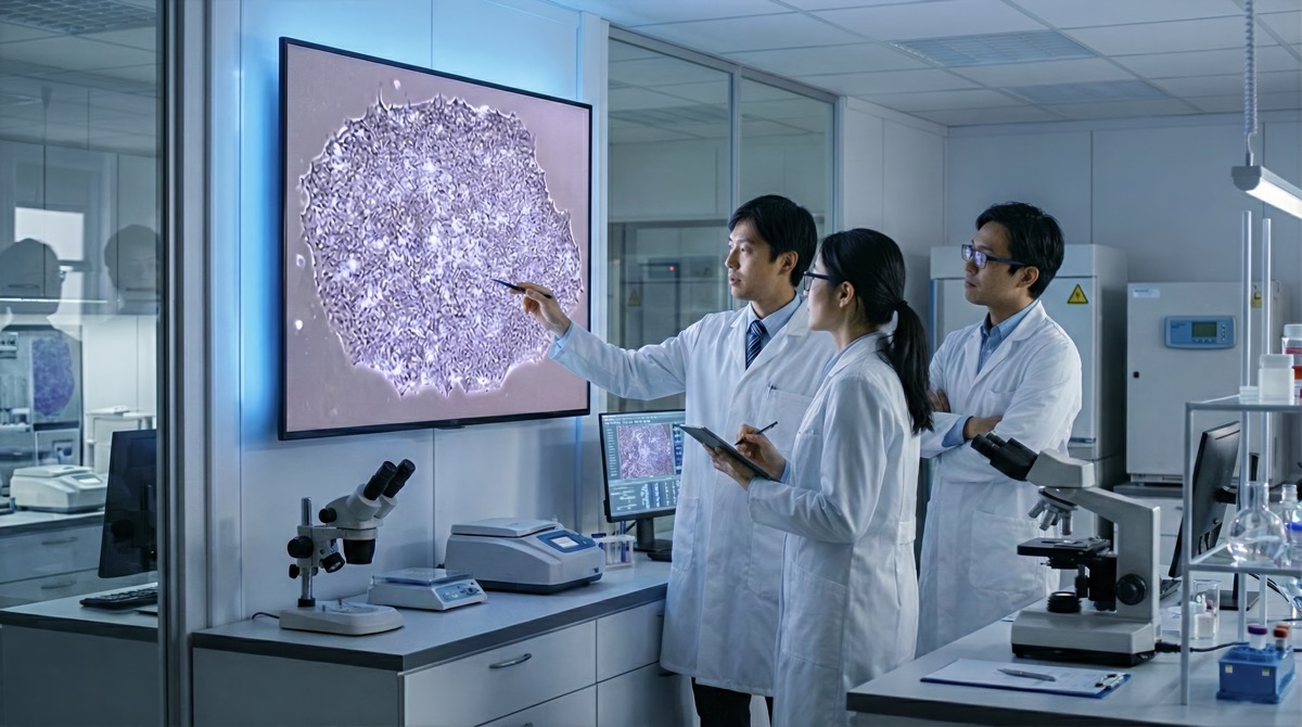
About the images:
The images in this article are AI-generated visuals created solely to illustrate the editorial content. They have been produced as original visual expressions and do not reproduce or derive from any third-party copyrighted material. No real individuals, protected artworks, or registered trademarks have been used without authorisation. These images are provided transparently, with full regard for applicable copyright and intellectual property rights.